


close
Choose Your Site
Global
Social Media
Views: 217 Author: Maiwei Biotech Publish Time: 2026-09-19 Origin: Site


Content Menu
● Injection Pressure vs. Contrast Flow Rate
● The Physics Behind Contrast Delivery
● Why Injection Pressure Matters in CT
>> CT Angiography Requires a Dense, Predictable Bolus
● How Pressure Influences Angiography Procedures
>> Key Differences Between CT and Angiography Injection
● High Pressure Does Not Automatically Mean Better Imaging
● Contrast Viscosity: The Hidden Variable
● A Practical Injection Pathway Checklist
>> Before Connecting the Patient
● Pressure Tubing and Connectors Are Part of Image Quality
● Safety Priorities for Pressure-Controlled Injection
● Selecting Reliable Contrast Injection Consumables
● FAQ
>> 1. What is the difference between injection pressure and flow rate?
>> 2. Why does a CT injector show high pressure during contrast injection?
>> 3. Can high injection pressure improve CT angiography image quality?
>> 4. Why are high-pressure connecting tubes important in CT and angiography?
>> 5. Does warming contrast media reduce injection pressure?
>> 6. Can every PICC, port, or central venous catheter be used for power injection?
>> 7. What should radiology staff do if a patient reports pain during injection?
Injection pressure is a critical but often misunderstood factor in contrast-enhanced CT and angiography. It does not independently determine image quality; instead, it works with flow rate, contrast viscosity, catheter size, tubing resistance, vascular access, contrast volume, and scan timing to shape how reliably contrast media reaches the target vessel.
For radiology teams, distributors, and imaging consumables buyers, the practical objective is not to use the highest possible pressure. It is to achieve the prescribed contrast media delivery profile while keeping every component in the injection pathway within its labeled pressure and flow limits. In CT angiography and interventional angiography, this balance directly affects vessel enhancement, workflow consistency, patient comfort, and the risk of extravasation or device failure.

Injection pressure and flow rate are related, but they are not interchangeable.
Flow rate is the volume of contrast delivered per unit of time, usually expressed in mL/s. It is a clinical protocol setting that strongly influences bolus density and vascular enhancement.
Injection pressure is the force required to move contrast media through the syringe, high-pressure connecting tube, catheter, vascular access device, and patient's vascular system. It is often displayed in psi by a high-pressure injector.
A CT or angiography injector may be programmed for a target flow rate, but the pressure required to achieve that flow rate changes from case to case.
| Factor | Effect on Required Injection Pressure | Why It Matters |
|---|---|---|
| Higher flow rate | Increases pressure | More contrast must move through the system in less time |
| Smaller catheter internal diameter | Increases pressure substantially | Small changes in lumen diameter can create major resistance |
| Longer connecting tube | Increases pressure | Longer fluid pathways create more resistance |
| More viscous contrast media | Increases pressure | Thick contrast media are harder to move at the same flow rate |
| Lower contrast temperature | Increases pressure | Cooler contrast is generally more viscous |
| Kinked tubing or catheter | Can sharply increase pressure | Obstruction may compromise delivery and increase safety risk |
| Narrow, fragile, or poorly positioned access | Can increase pressure | Vascular access is often the weakest point in the delivery chain |
| Saline flush after contrast | May improve bolus use | It can push residual contrast from the tubing into circulation |
The core principle is simple: the injector creates enough pressure to deliver the selected flow rate, but the complete system must be rated to safely tolerate that pressure.
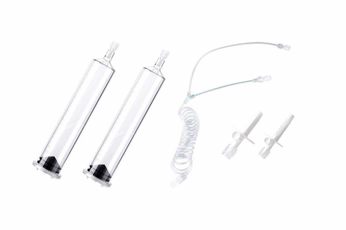
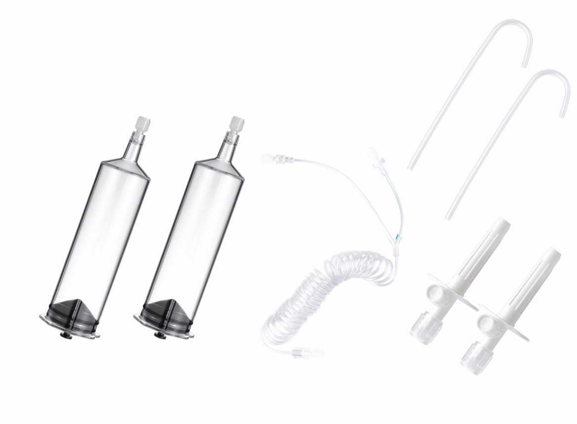
From a manufacturing perspective, this is why high-pressure connecting tubes, injector syringes, extension lines, and compatible accessories should be evaluated as a system rather than as unrelated disposable products. A pressure-rated tube may perform correctly in isolation, yet overall delivery can still be compromised by an incompatible connector, an unsuitable catheter, trapped air, an incorrect pressure setting, or improper assembly.
Contrast injection is governed by fluid resistance. In simplified terms, resistance rises when fluid becomes more viscous, the fluid path becomes longer, or the internal diameter becomes smaller.
For laminar flow through a cylindrical pathway, Poiseuille's law illustrates the relationship:
Q=πΔPr4/8ηL
Where:
- Q = flow rate
- ΔP = pressure difference
- r = internal radius of the tube or catheter
- η = fluid viscosity
- L = length of the pathway
The most important practical insight is the r4 relationship. A modest reduction in the internal radius of a catheter or tube can create a disproportionately large increase in resistance. This explains why a high-flow CT angiography protocol may work smoothly through a suitable large-bore peripheral IV line but trigger high-pressure alarms when used with a smaller-gauge catheter or a compromised access site.
In real clinical conditions, contrast delivery is more complex than this equation alone. The pathway includes connectors, valves, bends, pressure tubing, patient-specific venous anatomy, and changes in contrast temperature. Still, the principle remains valuable: when resistance rises, the system needs more pressure to maintain the same flow rate.
CT imaging frequently depends on a well-defined enhancement phase. For routine contrast-enhanced CT, the goal may be venous, portal venous, delayed, or multiphasic enhancement. For CT angiography, the requirement is often more demanding: contrast must arrive at the arterial target during a narrow scan window with sufficient iodine delivery to make vessels conspicuous.
A CT injector must therefore deliver a consistent bolus with the intended:
- Contrast volume
- Iodine concentration
- Flow rate
- Injection duration
- Saline chaser volume and flow
- Scan delay or bolus-tracking trigger
- Pressure limit
If pressure is too low for the resistance in the system, the programmed flow rate may not be achieved. The result can be reduced arterial opacification, an elongated or dispersed bolus, poor contrast timing, or a nondiagnostic CTA study.
If the pressure limit is too high relative to the rated capacity of the tubing, catheter, port, PICC, or connector, the risk profile changes. High pressure does not automatically improve enhancement. It may instead indicate resistance that requires investigation.
CT angiography is particularly sensitive to the relationship between flow rate and iodine delivery rate. A useful concept is:
Iodine Delivery Rate=Contrast Flow Rate×Iodine Concentration
For example, contrast at 370 mg iodine/mL delivered at 4 mL/s provides a higher iodine delivery rate than the same agent delivered at 2 mL/s. However, increasing the flow rate may also increase the pressure demand across the tubing and vascular access pathway.
For a CTA protocol, the imaging team should not simply increase pressure to solve inadequate enhancement. A systematic review is more appropriate:
1. Confirm the prescribed flow rate and contrast concentration.
2. Verify the IV catheter gauge, location, and patency.
3. Check the rated pressure and flow limits of the catheter and connecting tube.
4. Inspect the high-pressure tubing for kinks, loose connections, or air.
5. Confirm that contrast temperature and viscosity are appropriate for the protocol.
6. Review the saline chaser and bolus-tracking strategy.
7. Adjust the protocol only within departmental policies and device labeling.
This approach protects both image quality and patient safety.
In angiography, contrast media may be delivered through catheters positioned in arteries, veins, or selected vascular territories. The clinical purpose can include vessel mapping, lesion evaluation, embolization planning, stent placement, post-procedure assessment, or other image-guided interventions.
Unlike routine peripheral intravenous CT contrast injection, angiography may involve selective catheter placement and smaller internal lumens. These conditions can make pressure management especially important.
The injection system must generate sufficient pressure to deliver contrast at the desired rate through the catheter, while respecting the pressure rating of every component. A high-pressure angiography tubing set needs to provide a secure connection between the injector and the patient-side delivery device without compromising the intended flow profile.
| Consideration | CT Contrast Injection | Angiography Contrast Injection |
|---|---|---|
| Typical delivery route | Peripheral IV, power-injectable port, or approved central access | Selective angiographic catheter or vascular sheath |
| Main imaging objective | Timed enhancement of organs, veins, or arteries | Real-time visualization of specific vessels and lesions |
| Importance of bolus timing | Very high, especially in CTA | High, but often coordinated with live fluoroscopy or digital subtraction imaging |
| Pressure sensitivity | Influenced by IV gauge, access site, tubing, and viscosity | Influenced heavily by catheter lumen, catheter position, vascular territory, and injection protocol |
| Common workflow concern | Achieving diagnostic enhancement during the scan window | Maintaining controlled vascular opacification without exceeding device limits |
| Consumables focus | CT injector syringe, high-pressure CT tubing, extension line, check valve | Angiography injector tubing, high-pressure manifold components, catheter-compatible connectors |
The common requirement is controlled contrast delivery through a pressure-rated system. The practical differences come from access route, target anatomy, procedure type, and the delivery hardware used.
A common operational mistake is to interpret high injector pressure as a sign of "stronger" or "better" contrast delivery. In reality, pressure is often a signal of resistance.
When the injector reports unexpectedly high pressure, the clinical team should consider whether the pathway has become restrictive. Potential causes include:
- A small-gauge peripheral IV catheter used for a high-flow protocol
- A catheter placed in a distal hand vein rather than a suitable antecubital vein
- Partial infiltration or poor venous patency
- A kinked, compressed, or improperly routed connecting tube
- High-viscosity contrast media, especially if not warmed when appropriate
- A blocked connector, closed clamp, or defective valve
- An access device not labeled for power injection
- Flow rate settings that do not match the available vascular access
In other words, high pressure can be an operational warning, not a performance advantage.
For facilities that use multiple injector brands or work with various disposable sets, compatibility management is essential. A connecting tube should be designed and validated for the relevant injector interface, procedure type, pressure range, and intended contrast-delivery environment. Procurement teams should also verify traceability, lot control, sterility status, connector configuration, packaging integrity, and applicable regulatory documentation.
Viscosity is one of the most important variables affecting injection pressure. More viscous contrast media requires more force to achieve the same flow rate through the same catheter and tubing configuration.
Viscosity is affected by:
- Contrast formulation
- Iodine concentration
- Temperature
- Storage conditions
- Injection speed
- Catheter diameter
- Length of the delivery pathway
Warming iodinated contrast media can reduce viscosity, which may lower resistance and make high-flow injection more manageable. Guidance from European radiology experts notes that warming iodine-based contrast can reduce viscosity and may reduce the risk of contrast extravasation, although local protocols, product instructions, and clinical judgment must guide practice.
For manufacturers of CT, MRI, and angiography consumables, viscosity reinforces the need for robust high-pressure delivery components. The disposable set must support consistent fluid transmission under expected clinical conditions, including high-viscosity contrast, saline flushing, multiphase injection, and scanner-table movement.
The most reliable way to control injection pressure is to assess the complete delivery pathway before injection. The following checklist can help radiology teams standardize preparation.
- Confirm that the injector syringe and high-pressure connecting tube match the intended CT, MRI, or angiography application.
- Verify the sterile package integrity, expiration date, lot information, and product labeling.
- Check connector type and compatibility with the injector system and vascular access device.
- Ensure the entire fluid pathway is correctly assembled and secured.
- Remove air from syringes, connecting tubes, and patient-side components according to departmental procedure.
- Confirm that the programmed pressure limit does not exceed the rated limit of the tubing, catheter, port, PICC, or other vascular access device.
- Confirm contrast media type, concentration, volume, injection rate, and scan delay.
- Assess the selected vascular access site and catheter condition.
- Confirm blood return or use a saline test injection when required by local procedure.
- Check for tenderness, swelling, leakage, or signs of infiltration.
- Ensure sufficient tubing slack for scanner-table movement.
- Tell the patient what sensations to report, including pain, burning, swelling, tightness, or discomfort at the injection site.
- Monitor for unexpected pressure changes or injector alarms.
- Observe the injection site whenever feasible.
- Stop the injection and assess immediately if there is pain, swelling, resistance, or suspected extravasation.
- Document the injection protocol, access route, consumables used, and any incident according to local requirements.
- Replace any contaminated disposable component according to infection-control procedure.
A consistent checklist is not only a safety tool. It also improves repeatability across technologists, shifts, scanners, and sites.
It is easy to view injector tubing as a basic accessory. In practice, it is a functional component of the contrast-delivery system.
High-pressure connecting tubes and compatible connectors must reliably transmit the intended contrast volume, injection pressure, and flow rate from the injector to the patient. They also need to remain secure during patient positioning and scanner-table movement.
A suitable high-pressure tubing solution should support:
- Stable connection performance during injection
- Clear product labeling for intended use and pressure rating
- Compatibility options for commonly used injector systems where applicable
- Reliable manufacturing consistency across production batches
- Sterile, single-use configurations when required
- Controlled internal pathway design to support predictable fluid delivery
- Appropriate traceability and quality documentation
- OEM customization capability for distributors and device partners when needed
For MAIWEI INC., the value proposition is not simply supplying a disposable accessory. It is supporting a more dependable contrast-delivery workflow for CT, MRI, and angiography environments through compatible high-pressure injectors, connecting tubes, and related consumables.
Pressure-controlled injection should always be managed according to the specific injector instructions, contrast media instructions, vascular access labeling, institutional protocol, and the clinical judgment of qualified healthcare professionals.
Several practical safety principles are consistently important:
- Use only vascular access devices approved for the intended injection conditions.
- Never assume that a central venous catheter, PICC, port, or extension line is suitable for power injection without checking its labeling.
- Match catheter size and access location to the planned flow rate.
- Do not use conventional low-pressure IV tubing as a substitute for high-pressure injector tubing.
- Treat unexpected resistance as a reason to assess the pathway, not simply to raise the pressure limit.
- Use a careful saline test injection and visual site monitoring where appropriate.
- Minimize air in the system through standardized setup procedures.
- Use the lowest contrast dose and most appropriate protocol that still provides a diagnostic examination.
A clinical review of pressure injectors emphasizes that pressure tubing and connectors are required to accurately transmit selected volume, pressure, flow, and rate between the injector system and the patient. It also highlights the importance of clearing air, checking vascular access, allowing tubing slack during table movement, and maintaining patient communication.
Hospitals, distributors, and OEM buyers should evaluate contrast injection consumables beyond unit price. Poorly matched or inconsistently manufactured components can create operational interruptions, unnecessary waste, and avoidable risk.
When evaluating a CT injector tubing manufacturer or angiography consumables supplier, consider the following questions:
1. Is the product designed for the intended imaging modality and injector configuration?
2. Is the pressure rating clearly defined and aligned with the planned clinical use?
3. Is compatibility supported by product documentation and appropriate validation?
4. Does the supplier maintain a medical-device quality management system?
5. Are sterility, biocompatibility, packaging, labeling, and traceability controls documented?
6. Can the supplier support stable global delivery, OEM branding, and regulatory documentation?
7. Are samples, technical specifications, and quality records available for evaluation?
MAIWEI INC. develops and manufactures consumables for CT, MRI, and angiography high-pressure injector applications, including injectors, connecting tubes, and supporting disposable products compatible with multiple mainstream injection systems. With ISO 13485, CE, and FDA 510(k)-related product compliance capabilities, the company supports hospitals, distributors, and OEM partners in more than 60 countries and regions with medical imaging consumables designed for dependable workflow integration.
Injection pressure is not a standalone setting to maximize. It is the system response required to deliver contrast media at the planned flow rate through a specific pathway.
In CT, appropriate pressure supports the precise bolus needed for diagnostic enhancement and scan timing. In angiography, it helps deliver controlled contrast opacification through selective catheters and vascular access devices. In both settings, the safest and most effective result comes from matching the injector protocol, contrast properties, tubing set, connector design, vascular access, and pressure limit.
For imaging departments and supply partners, the right high-pressure injection consumables help turn a technically correct protocol into a repeatable clinical workflow.
Looking for compatible CT, MRI, or angiography high-pressure injection consumables? Contact MAIWEI INC. to discuss injector compatibility, high-pressure connecting tube configurations, OEM requirements, quality documentation, and sample evaluation for your market.
Flow rate is the amount of contrast media delivered each second, usually measured in mL/s. Injection pressure is the force the injector must generate to achieve that flow through the tubing, catheter, and vascular access pathway. Higher flow rates usually require higher pressure, but resistance from viscosity, catheter size, and tubing length also affects pressure.
High pressure usually means the injector is encountering resistance. Possible causes include a small catheter, high-viscosity contrast, cold contrast media, a narrow or poorly positioned IV, kinked tubing, a partially obstructed connector, or a programmed flow rate that is too high for the selected access device.
Not by itself. CTA quality depends on adequate iodine delivery, flow rate, injection duration, scan timing, patient circulation, and proper vascular access. High pressure may be necessary to maintain the planned flow rate, but an unexpectedly high pressure reading should prompt an assessment of the injection pathway.
High-pressure connecting tubes are designed to transfer contrast media from the injector to the patient-side access device under the intended pressure conditions. They are an important part of maintaining a secure, consistent delivery pathway and should be selected according to the injector system, procedure type, pressure rating, and compatibility requirements.
Warming iodinated contrast media generally reduces viscosity. Lower viscosity can reduce resistance in the fluid pathway and may help support high-flow injection. The decision to warm contrast should follow local procedures, product instructions, and clinical requirements.
No. Only devices specifically labeled for power injection should be used under the stated pressure and flow conditions. The device labeling should be checked before injection because conventional central lines and non-power-injectable access devices may not tolerate the pressures generated by CT or MRI injectors.
The injection should be stopped immediately and the site assessed according to institutional procedure. Pain, burning, swelling, tightness, or leakage may indicate extravasation or another access-related issue. The event should be managed and documented according to the facility's clinical protocol.
1. American College of Radiology. [Manual on Contrast Media].
2. Indrajit IK, Sivasankar R, D'Souza J, et al. [Pressure Injectors for Radiologists: A Review and What Is New]. *Indian Journal of Radiology and Imaging*. 2015;25(1):2–10.
3. European Society of Urogenital Radiology. [ESUR Guidelines on Contrast Agents, Version 10.0].
4. Roditi G, Khan N, van der Molen AJ, et al. [Intravenous Contrast Medium Extravasation: Systematic Review and Updated ESUR Contrast Media Safety Committee Guidelines]. *European Radiology*. 2022.
5. UCSF Radiology. [Vascular Access and Use of Central Lines and Ports in Adults].
6. U.S. Food and Drug Administration. [Safety Considerations for Vascular Access Devices Used With Power Injectors].