


close
Choose Your Site
Global
Social Media
Views: 237 Author: Maiwei Biotech Publish Time: 2026-10-28 Origin: Site


Content Menu
● CT Injector Syringe Leakage vs. Contrast Extravasation
● What Causes CT Injector Syringe Leakage?
>> Loose, Misaligned, or Overtightened Connections
>> Incorrect Syringe Loading or Plunger Engagement
>> Damaged Syringe Barrels, Outlets, or Seals
>> Incompatible Syringes and Connecting Tubes
>> Excessive Resistance in the Fluid Pathway
>> Tubing Tension During Table Movement
● How Pressure, Flow Rate, and Contrast Viscosity Interact
>> Can Contrast Warming Prevent Leakage?
● A Practical Checklist to Prevent CT Injector Syringe Leakage
>> 1. Verify the Exact Consumable Configuration
>> 2. Inspect Packaging and Components
>> 3. Load and Connect According to Instructions
>> 4. Prime the Pathway and Remove Air
>> 5. Confirm Vascular Access Suitability
>> 6. Check Routing and Monitor the Injection
● What Should Staff Do When a Leak Occurs?
● Investigating Recurring Leakage: Follow the Pattern
>> Use a Meaningful Denominator
● What Buyers Should Request From a Consumables Manufacturer
>> MAIWEI INC.'s Approach to Product Selection
● Build Leakage Prevention Into Every Injection
>> Why Does a CT Injector Syringe Leak Only During Injection?
>> Can Overtightening Stop a Leaking Connector?
>> Is a Higher Pressure Limit a Solution to Leakage?
>> Can a Leaking Single-Use Syringe Be Reused?
>> Are CT, MRI, and Angiography Syringes Interchangeable?
>> What Should Be Included in a Leakage Complaint?
CT injector syringe leakage can result from loose connections, damaged syringe components, incompatible consumables, incorrect loading, or pressure that exceeds a component's rated operating limit. Preventing it requires more than tightening a connector: the entire fluid pathway must be correctly selected, assembled, inspected, and monitored.
For hospitals, distributors, and equipment partners, the most useful approach is to treat the syringe, connecting tube, catheter, and injector as one system. From our manufacturing perspective at MAIWEI INC., compatibility should mean documented suitability for a particular application—not simply that two components appear to fit.
This guide explains how to recognize different leakage patterns, reduce preventable failures, and investigate recurring problems. It supports trained professionals; device instructions for use and institutional clinical protocols always take precedence.

Before investigating a suspected leak, identify where the fluid is escaping.
External leakage occurs outside the patient. Contrast or saline may appear around the syringe outlet, behind the plunger, along the connecting tube, or at a connector.
Contrast extravasation occurs when injected fluid escapes the vascular space into surrounding tissue. The syringe and tubing may remain completely dry.
These are different events, although both can interrupt an examination and require immediate assessment.
| Observation | Possible issue | Immediate priority |
|---|---|---|
| Fluid around the syringe outlet | Loose, damaged, or incorrectly assembled connection | Stop injection and inspect the fluid pathway |
| Moisture behind the syringe plunger | Possible seal or syringe damage | Remove the affected disposable from service |
| Fluid along the connecting tube | Tubing damage or connector failure | Replace affected components |
| Pain, swelling, or tightness at the injection site | Possible extravasation | Stop injection and initiate clinical assessment |
| Pressure alarm without visible fluid | Obstruction, resistance, loading problem, or another system fault | Follow the injector's troubleshooting instructions |
A dry syringe does not rule out extravasation. Likewise, fluid near the injector does not prove that the syringe barrel itself failed.
An incompletely secured connection may leak when injection pressure rises. Misalignment can also prevent proper engagement.
However, applying more force is not automatically the solution. Excessive tightening can damage plastic connectors or threads.
Staff should assemble connections using the method specified by the manufacturer. Do not use tools, tape, adhesives, or improvised sealing materials unless explicitly authorized in the applicable instructions.
A syringe may appear installed without being fully seated or correctly engaged with the injector piston.
Depending on the system, incorrect loading can interfere with plunger movement or injection performance. The response should be to verify installation—not to force the syringe into position.
Check the retaining mechanism, syringe orientation, and plunger engagement according to the injector manual. Any loading uncertainty should be resolved before patient connection.
Cracks, deformation, or damage around the outlet and plunger seal can compromise fluid containment.
Possible contributing factors include handling damage, unsuitable storage, or manufacturing defects. Visual inspection is essential, but it cannot establish that every component will perform correctly under pressure.
Reject products with damaged packaging, visible cracks, displaced components, or evidence of leakage. Follow labeled storage conditions and expiry dates.
Physical fit does not establish pressure-injection compatibility.
Two syringes may have similar capacities but differ in barrel geometry, plunger engagement, retention features, or validated operating conditions. Similarly, a connector that attaches successfully may not be appropriate for the intended pressure and flow.
Verify the exact injector model and disposable part number. Do not assume that CT, MRI, and angiography consumables are interchangeable.
Kinked tubing, closed clamps, unsuitable catheters, and other restrictions can increase resistance.
Contrast viscosity also affects injection pressure. More viscous contrast generally requires greater pressure to achieve a given flow through the same pathway.
A pressure-limit alarm is not an instruction to raise the limit. Identify the cause and confirm that the prescribed protocol remains appropriate.
A connection that remains stable during setup may experience additional stress when the patient table moves.
Tubing caught under equipment, stretched across the table, or routed around sharp bends can create avoidable problems.
Provide sufficient slack for planned movement while keeping the pathway visible and protected from snagging.
A useful troubleshooting principle is that flow rate and pressure are related, but they are not interchangeable.
The pressure required to deliver a selected flow depends on factors including:
- Contrast viscosity.
- Tubing length and internal diameter.
- Catheter characteristics.
- Connectors and other restrictions.
- Kinks, closed clamps, or partial obstructions.
The permitted operating conditions must respect every component's labeling. A syringe's pressure rating does not authorize the same pressure through an inadequately rated extension tube or vascular access device.
Warming iodinated contrast under an approved protocol can reduce viscosity and the pressure needed for injection.
However, warming cannot repair a cracked syringe, correct incompatible components, or guarantee prevention of leakage or extravasation.
Use only the contrast product's permitted warming and storage conditions. Do not apply iodinated-contrast practices automatically to MRI contrast agents.
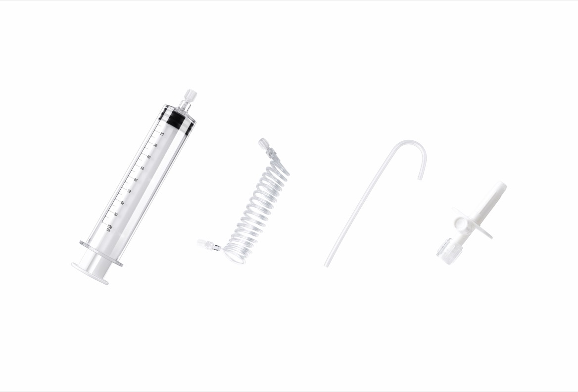
A consistent preparation sequence helps teams avoid relying on memory during busy lists.
Confirm the injector model, syringe reference, connecting tube, and intended protocol.
Check documented compatibility and applicable pressure and flow limits. Resolve discrepancies before opening the sterile package.
Check package integrity, expiry date, and visible component condition.
Look for cracks, damaged threads, displaced seals, distorted tubing, and unexplained moisture. Do not use a questionable disposable to see whether it "holds."
Seat the syringe correctly and verify plunger engagement.
Assemble connectors without cross-threading or excessive force. Maintain aseptic technique throughout preparation.
Remove air from the syringe and associated tubing using the approved procedure.
Keep the patient disconnected during purging and other operations for which the injector instructions require disconnection. Never bypass an air-check step to save time.
Qualified clinical staff should assess access suitability for the planned injection.
A saline assessment may form part of the institutional protocol, but it does not guarantee that a later contrast injection will be complication-free.
Confirm that tubing remains free of kinks and tension throughout expected table movement.
Monitor the patient, injection site, equipment, and injector feedback according to local practice. Encourage patients to report pain, burning, swelling, or unusual pressure immediately.
Stop the injection promptly and follow the device instructions and institutional response procedure.
The priorities are patient assessment, safe control of the fluid pathway, and preservation of information needed to understand the event.
1. Assess the patient and injection site for possible extravasation.
2. Identify visible leakage without exposing staff or the patient to additional risk.
3. Remove damaged or suspect disposables from service.
4. Preserve product identifiers and affected items where safe and permitted.
5. Document the event and escalate through clinical, biomedical, and quality channels.
6. Resume only after appropriate reassessment and correction.
Do not attempt to repair a leaking disposable while it remains connected to the patient.
If delivered contrast volume or bolus timing is uncertain, the responsible clinician should assess examination adequacy. Repeating an injection is not an automatic troubleshooting step.
Repeated failures deserve a structured investigation rather than an assumption of "operator error" or "bad syringes."
Useful questions include:
- Does the problem follow one product lot?
- Does it occur on one injector or several?
- Is the leakage always at the same connection?
- Did a tubing supplier or protocol recently change?
- Does it occur only at particular flow settings?
- Was table movement involved?
Record the injector identifier, disposable references, lot numbers, contrast product, programmed settings, visible failure location, and relevant alarm information.
Ten complaints do not establish a failure rate unless the number of products used is known.
A practical internal metric is confirmed external-leakage events per 1,000 uses of a defined configuration. Track suspected events separately until investigation clarifies them.
Do not combine syringe leakage, tubing disconnection, and patient extravasation into one undifferentiated category. They require different corrective actions.
Published adverse-event reports can help identify possible failure modes, but they do not independently establish incidence or prove causation.
For procurement teams, a "high-pressure" label is only the beginning.
Request documentation covering:
| Evaluation area | Evidence to request |
|---|---|
| Compatibility | Exact injector models and supported product references |
| Operating limits | Labeled pressure, flow, and use conditions |
| Performance | Relevant leakage, connection, and integrity test evidence |
| Sterile supply | Sterilization information, packaging integrity, and shelf life |
| Traceability | Lot identification and complaint-investigation procedures |
| Regulatory status | Documentation applicable to the exact product and market |
Ask how performance was evaluated in the intended configuration—not simply whether an individual syringe passed a standalone test.
ISO 13485 certification concerns the quality management system. It does not mean every product is automatically authorized in every market. Likewise, FDA 510(k) clearance is device-specific and should not be described as blanket company approval.
MAIWEI INC. develops and manufactures high-pressure injector syringes, connecting tubes, and related consumables for CT, MRI, and angiography applications.
Our company profile reports supply to more than 60 countries and regions, with ISO 13485 certification and relevant CE and FDA 510(k) documentation. Buyers should confirm the scope and applicability of these documents for each selected product.
For hospitals, distributors, and OEM partners, the practical starting point is the same: identify the intended equipment and application before selecting consumables.
Reliable prevention combines suitable consumables, correct assembly, controlled operating conditions, and attentive monitoring.
Contact MAIWEI INC. with your injector model, required syringe capacity, connecting-tube configuration, and target market. Request product-specific compatibility information, applicable regulatory documents, and samples for evaluation under your institution's procedures.
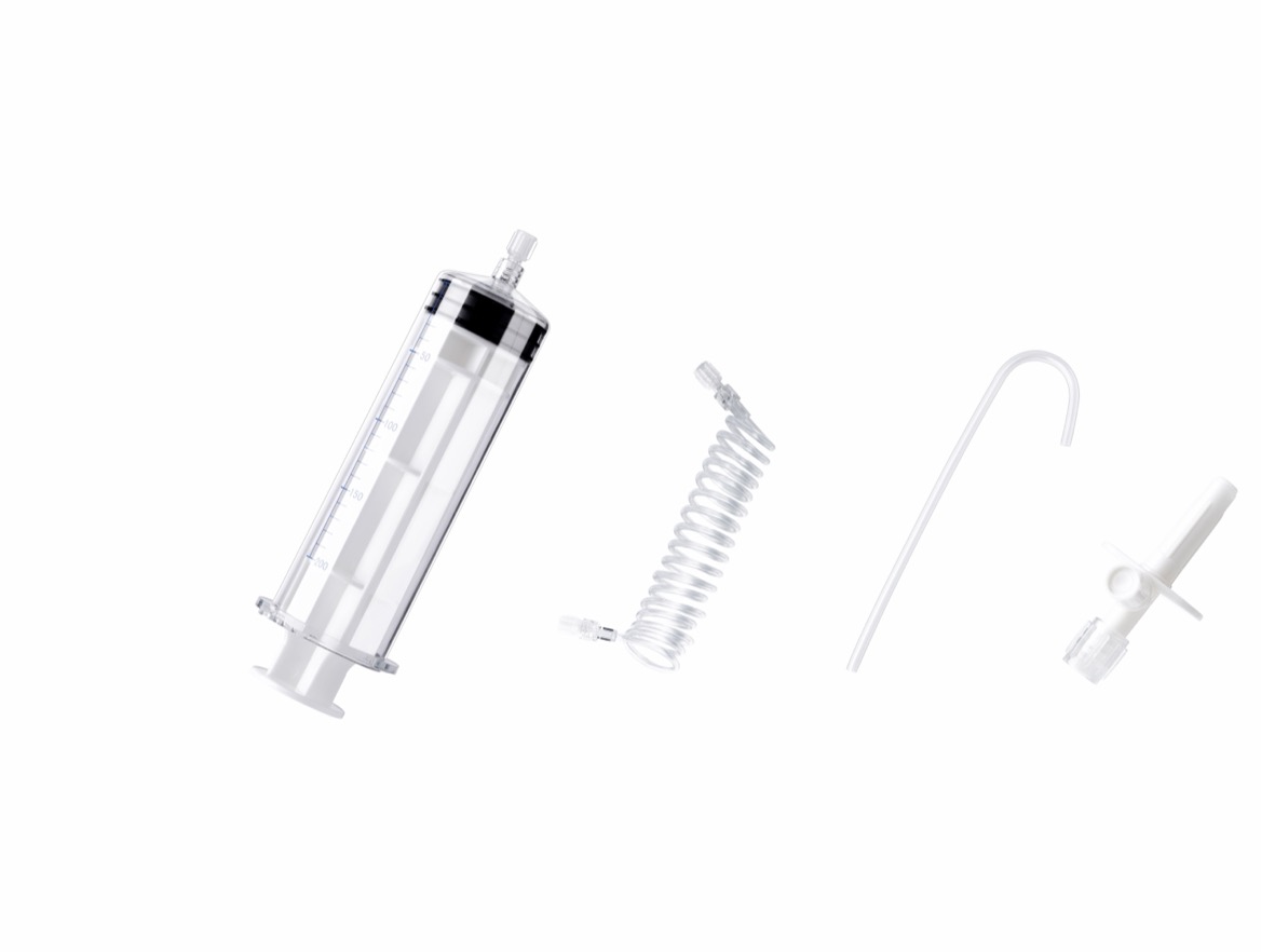
A marginal connection or damaged component may remain dry at rest but leak as pressure rises. Stop use and investigate the complete pathway rather than assuming the syringe alone caused the problem.
Not reliably. Excessive tightening can damage plastic components. Follow the specified assembly method and replace suspect parts instead of applying additional force.
No. Raising the limit does not correct damage or incompatibility. It may expose components to conditions outside their labeling. Investigate resistance and verify all operating limits.
No. Remove the affected syringe from service. Do not patch, reprocess, or reuse a product labeled for single use.
Not automatically. Requirements differ by equipment and application. Use only documented compatible configurations within their specified conditions.
Include product references, lot numbers, injector model, injection settings, failure location, photographs where permitted, and patient impact. Preserve affected components according to institutional procedures and manufacturer guidance.
1. American College of Radiology. *ACR Manual on Contrast Media*. Professional guidance covering contrast administration, extravasation, and contrast warming. [acr]
2. Bayer HealthCare. *MEDRAD Stellant Operation Manual*, available through ManualsLib. Equipment-specific instructions covering syringe installation, compatible pressure ratings, air removal, and patient-disconnection precautions. Consult the current manufacturer-issued manual for the exact system in use. [manualslib]
3. *Pressure Injectors for Radiologists: A Review and What Is New*. Peer-reviewed review available through PubMed Central. Discusses pressure-injector components, preparation procedures, tubing management, and injection safety. [pmc.ncbi.nlm.nih]
4. *Intravenous Contrast Medium Extravasation: Systematic Review and Updated ESUR Contrast Media Safety Committee Guidelines*. Peer-reviewed guidance published in 2022, available through PubMed Central. Addresses extravasation risk factors, contrast viscosity, warming, and clinical management. [pmc.ncbi.nlm.nih]
5. Royal Australian and New Zealand College of Radiologists. *Iodinated Contrast Media Guideline*, Version 2.3, March 2018. Guidance on vascular access assessment, injection-site monitoring, contrast viscosity, and warming. [ranzcr]
6. U.S. Food and Drug Administration. *Device Approvals and Clearances*. Explains the distinction between device clearance through the 510(k) pathway and premarket approval. [fda]
7. U.S. Food and Drug Administration. *Contrast and Saline Syringe Prescribing Information*, 2012 labeling document. Includes inspection, leakage rejection, aseptic handling, and air-removal precautions for the labeled products. [accessdata.fda]