


close
Choose Your Site
Global
Social Media
Views: 255 Author: Maiwei Biotech Publish Time: 2026-10-13 Origin: Site


Content Menu
● Contrast Injector Syringe Leakage vs. Contrast Extravasation
● Immediate Steps When a Contrast Injector Syringe Leaks
>> 1. Stop Injection and Alert the Team
>> 2. Assess the Patient Before Troubleshooting
>> 3. Make the System Safe Before Handling Connections
>> 4. Inspect the Entire Fluid Pathway
>> 5. Replace Compromised Components and Reassess
● Why Does a High-Pressure Injector Syringe Start Leaking?
>> Incorrect Assembly or Connector Damage
>> Syringe or Plunger-Seal Problems
>> Pressure or Flow Outside Component Limits
>> Resistance Elsewhere in the Pathway
● Can I Tighten the Connection or Lower the Injection Rate?
● What to Do If Contrast Extravasation Is Suspected
>> Warning Signs Requiring Urgent Escalation
● What Published Data Reveal—and What They Do Not
>> A Documented Industry Safety Lesson
● A Practical Root-Cause Investigation After Leakage
● Preventing Contrast Injector Syringe Leakage
>> Use a Complete-System Compatibility Check
>> Standardize Preparation and Observation
>> Track Equipment Leakage Separately
● Choosing a Reliable Imaging Consumables Partner
>> Can a Small Syringe Leak Be Ignored?
>> Does Syringe Leakage Always Mean Extravasation?
>> Can I Restart After Replacing the Syringe?
>> Will a Pressure Alarm Detect Every Leak?
>> Can CT, MRI, and Angiography Consumables Be Interchanged?
>> Should Every Extravasation Receive Surgical Consultation?
If a contrast injector syringe starts leaking during injection, stop the injection immediately, assess the patient, and notify the supervising clinical team. Do not tighten a connection while the system is pressurized or restart simply because the leak appears small. Follow the injector's instructions for safely relieving pressure before inspecting or replacing affected components.
Two problems must be distinguished: contrast escaping from the syringe or connecting tubing, and contrast leaking from the blood vessel into surrounding tissue. The second is called contrast extravasation. These events require different investigations, but both begin with stopping injection and protecting the patient.
From a manufacturer's perspective, troubleshooting should examine the complete fluid pathway—not just the visibly wet component. This guide explains the differences, practical response steps, and compatibility checks for trained imaging professionals working with CT, MRI, and angiography systems.
This educational article does not replace device instructions, institutional procedures, or clinical judgment.
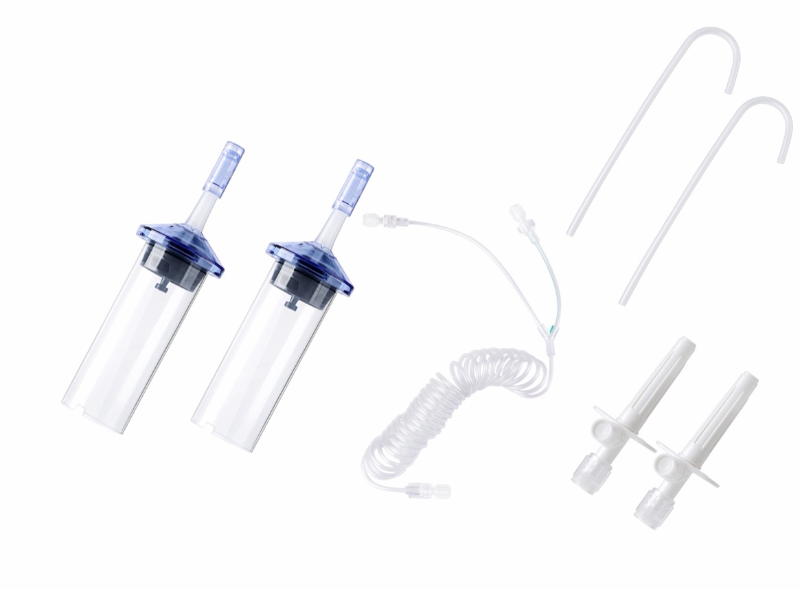
A wet syringe connection does not automatically indicate extravasation. Conversely, a dry injector does not prove that contrast is entering the vessel correctly.
| Comparison point | Syringe or tubing leakage | Contrast extravasation |
|---|---|---|
| Where contrast escapes | Outside the fluid pathway, at a syringe, connector, or tubing segment | Outside the blood vessel, into surrounding tissue |
| Typical observations | Visible droplets, wet connections, spraying, or damaged tubing | Pain, swelling, tightness, or changes around the access site |
| Main investigation | Component integrity, assembly, compatibility, and pressure limits | Vascular access and the patient's clinical condition |
| Immediate response | Stop injection and assess the patient before inspecting equipment | Stop injection and obtain prompt clinical assessment |
| Restart decision | Requires replacement or correction, verification, and clinical authorization | Depends on assessment, access management, and the imaging plan |
Both problems may occur during the same interrupted examination. Inspecting the equipment must never delay patient assessment.
In angiography, leakage near arterial access or an invasive catheter requires the procedural team's immediate attention. Peripheral intravenous extravasation guidance should not be treated as a complete protocol for arterial complications.
Use the injector's designated stop control. Inform the supervising radiologist or procedural clinician and coordinate the interrupted examination.
Do not continue to "finish the bolus." Continued injection through a compromised fluid pathway may increase fluid loss, exposure, or patient injury.
If the patient develops breathing difficulty, collapse, or another acute systemic symptom, activate the appropriate emergency response. A visible equipment leak does not exclude a separate contrast reaction.
Check the vascular access site and ask about:
- Pain, burning, or pressure.
- Swelling or tightness.
- Numbness or altered sensation.
- Difficulty moving the affected hand or limb.
The responsible clinician should evaluate suspected extravasation and determine observation, treatment, or escalation.
Patients who cannot communicate reliably need particularly careful observation. Do not rely only on the absence of reported pain.
Follow the model-specific instructions for stopping operation and relieving residual pressure.
Do not disconnect, retighten, or manipulate a pressurized fitting. Protect staff and the patient from splashes, and manage any blood or fluid exposure under institutional procedures.
In MRI, maintain the department's magnetic-field safety requirements during troubleshooting. Do not bring unsuitable replacement equipment or tools into the scanner environment.
After the system is safe, inspect:
- The syringe barrel, outlet, and plunger-seal area.
- Syringe seating and injector engagement.
- High-pressure tubing and connector interfaces.
- Any adapters, valves, or extension components.
- The patient-side connection and vascular access device.
Document where fluid first appeared. Contrast can spread along tubing, making the wettest point different from the original leak location.
Replace damaged, suspect, or contamination-exposed disposable components according to their instructions and facility policy.
Before any further injection, trained staff must verify assembly, priming, air removal, vascular access, and appropriate operating settings.
The supervising clinician should decide whether the examination can continue, needs a revised protocol, or should be postponed.
Incomplete engagement, cross-threading, damaged interfaces, or excessive tightening are possible causes to investigate.
A fitting that appears connected may still be incorrectly assembled. Follow the specified connection method rather than applying additional force.
Do not use tape, adhesive, improvised seals, or unapproved adapters to repair a leaking disposable.
Fluid near the plunger may indicate a seal-related problem. Leakage from the barrel may indicate physical damage. Neither observation, by itself, proves a manufacturing defect.
Investigators should also examine loading, injector engagement, compatibility, handling, and storage conditions.
A useful quality investigation preserves these competing explanations until evidence identifies the cause.
The syringe is only one part of a pressure-bearing system. Tubing, connectors, vascular access devices, and other components also have labeled limits.
The injector's pressure setting must not exceed applicable component limits. Flow-rate restrictions must also be respected.
A syringe with a suitable pressure rating does not make an ordinary infusion extension set suitable for power injection.
Kinked tubing, a closed valve, restricted access, or other obstruction can increase resistance.
Contrast viscosity and fluid-path geometry also influence the pressure needed to deliver a selected flow rate.
However, leakage alone does not establish that excessive pressure caused the event. Review recorded operating data alongside physical findings.
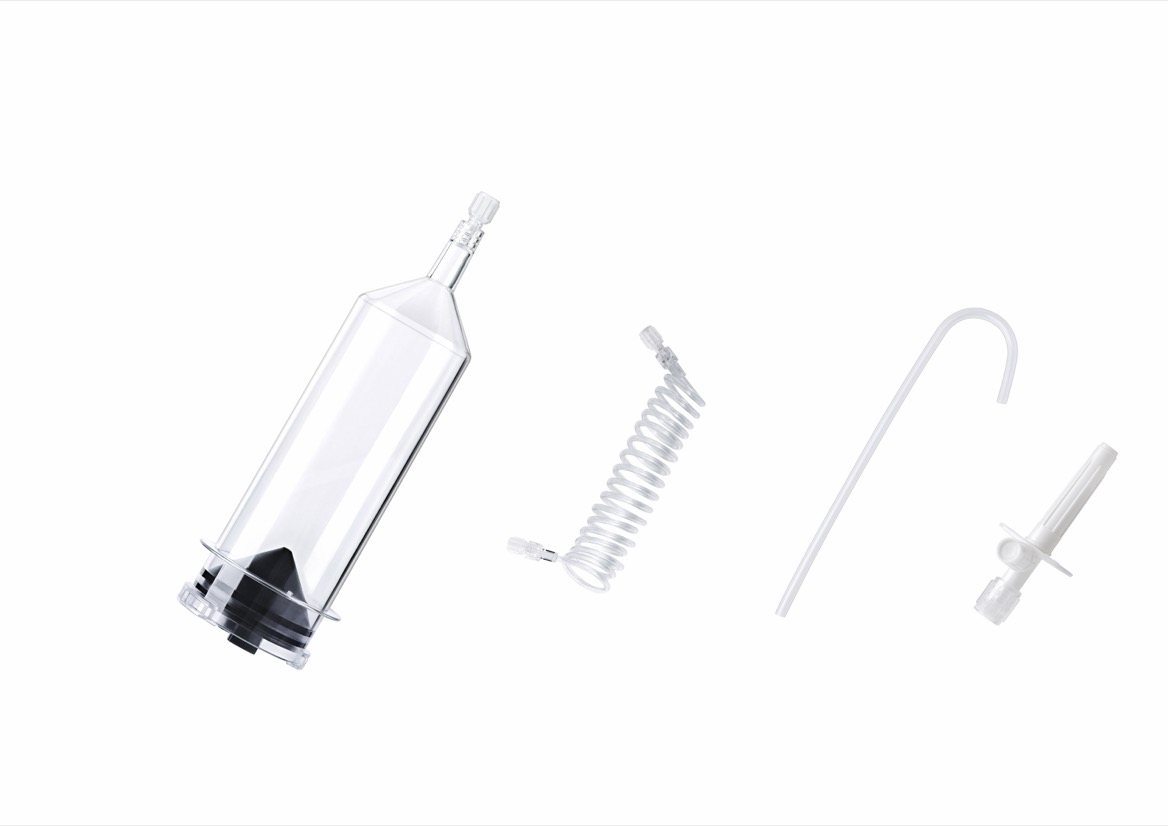
Neither action should be treated as an automatic solution.
| Proposed action | Why it is insufficient | Safer approach |
|---|---|---|
| Tighten during injection | The fitting remains pressurized and may be damaged | Stop and make the system safe first |
| Reduce the flow rate | A compromised seal or component may still leak | Identify the cause and replace suspect parts |
| Wipe away contrast and continue | Visible cleanup does not restore integrity or sterility | Reassess the fluid pathway |
| Reuse the damaged syringe | Damage may persist despite an apparently successful test | Use an appropriate replacement |
| Substitute a similar-looking component | Physical fit does not establish compatibility | Verify the exact product and injector combination |
Reducing flow or changing pressure settings may be part of an authorized revised protocol. It is not a repair for a defective or damaged component.
The clinical team should follow its approved extravasation protocol. Common measures include prompt assessment, limb elevation where appropriate, and locally prescribed cold-compress care.
Catheter management and any aspiration decision belong to trained clinicians following their institutional protocol. They are not universal do-it-yourself troubleshooting steps.
Seek urgent clinical escalation for:
- Severe or progressively worsening pain.
- Increasing swelling.
- Reduced perfusion.
- New sensory changes.
- Worsening movement.
- Skin blistering or ulceration.
Clinical severity is central to escalation decisions. A small estimated volume should not override concerning symptoms.
Patients discharged after assessment need clear instructions about worsening symptoms and where to obtain further care.
A 2022 systematic review and guideline update reported that an observational study of 142,651 CT examinations found an extravasation incidence of 0.23%. It also summarized a systematic review covering 1,104,872 patients with an incidence of approximately 0.2%.
These figures describe contrast extravasation, not external syringe leakage. They should not be presented as syringe failure rates or used to compare consumable manufacturers.
The distinction matters commercially and clinically. Combining equipment leaks with vascular extravasation produces misleading quality metrics.
A Pennsylvania patient-safety advisory described 29 reports of IV tubing rupture during contrast injection between July 2004 and March 2008.
Reported consequences included splashing patients and staff and interrupting examinations. The advisory highlighted the danger of using conventional IV sets instead of appropriate high-pressure components.
This historical reporting does not establish today's incidence. It demonstrates why compatibility must cover the complete connected system.
From a manufacturing-quality perspective, a useful incident report should answer three questions: where did the leak occur, under what conditions, and what evidence remains?
Record the following when available:
| Information | Investigation value |
|---|---|
| Injector model and identification | Establishes the equipment configuration |
| Syringe and tubing product codes | Identifies the exact components |
| Lot numbers and expiry dates | Supports traceability |
| Programmed flow and pressure limit | Describes intended operating conditions |
| Available pressure trace and alarms | Helps reconstruct the event |
| Contrast type and preparation conditions | Adds relevant fluid information |
| Photographs and leak location | Preserves visible findings |
| Patient assessment and examination outcome | Documents clinical consequences |
Retain suspect components under appropriate contamination-control procedures. Do not perform informal pressure testing on the patient or reconnect a suspect disposable to reproduce the fault.
Repeated events involving the same configuration warrant review by clinical engineering, quality personnel, and the supplier. Applicable incident-reporting obligations should also be considered.
Before procurement or clinical use, verify:
1. The exact injector model.
2. The specified syringe product.
3. The connecting-tube configuration.
4. Applicable pressure and flow limits.
5. Vascular access suitability.
6. Modality-specific requirements.
7. Current instructions and regulatory documentation.
"Compatible with major brands" is not enough for an individual purchasing decision.
A practical preparation checklist should include:
- Inspecting packaging, expiry, and visible component condition.
- Loading and connecting components as instructed.
- Priming and removing air according to the device instructions.
- Checking access under the approved clinical protocol.
- Positioning tubing to avoid kinks and unnecessary traction.
- Asking patients to report pain or swelling immediately.
An injector alarm is useful information, not a substitute for observing the patient and access site.
For internal quality improvement, distinguish external fluid-path leaks, tubing ruptures, access-site extravasation, and assembly-related incidents.
This proposed classification helps teams identify whether corrective action belongs in product selection, staff preparation, equipment maintenance, or vascular-access practice.
Avoid assigning blame before investigating the evidence.
MAIWEI INC. develops and manufactures high-pressure injector syringes, connecting tubes, and related consumables for CT, MRI, and angiography applications.
Based on company-provided information, MAIWEI serves hospitals, distributors, and OEM customers across more than 60 countries and regions.
For purchasing decisions, request documentation for the specific product and destination market. ISO 13485 concerns quality-management systems; CE status and FDA 510(k) clearance must be checked for their applicable product scope. They do not establish universal injector compatibility or guarantee leak-free use.
Contact MAIWEI with your injector model, required syringe and tubing configuration, operating requirements, and target market. Request a model-specific compatibility review, current instructions, and applicable regulatory documents before ordering samples or placing an OEM order.
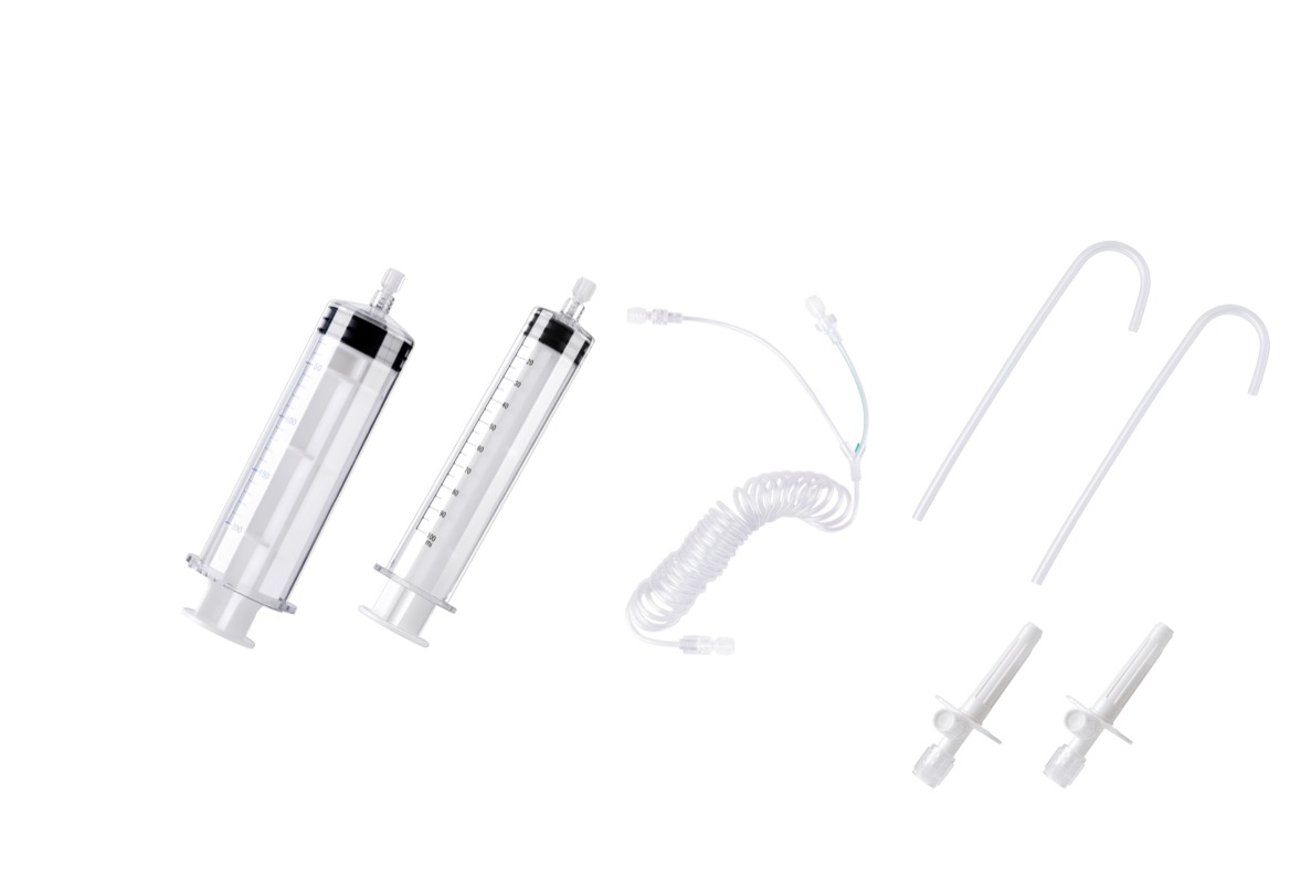
No. Stop injection and assess the patient. A small visible leak does not confirm that the fluid pathway remains safe or that the intended dose reached the circulation.
No. External leakage occurs at the equipment. Extravasation occurs outside the blood vessel. Inspecting both the equipment and the access site helps distinguish them.
Not automatically. The complete pathway, vascular access, delivered contrast, and imaging requirements need reassessment. Restart only after required checks and clinical authorization.
Do not assume so. Pressure monitoring provides useful information, but direct observation and patient communication remain important for recognizing problems.
Only when the specific combination is supported by the applicable instructions. Similar appearance or connector fit does not establish pressure, operational, or modality compatibility.
Not necessarily. The responsible clinician evaluates severity. Progressive pain, sensory changes, perfusion problems, or skin injury require urgent escalation; volume alone is not an adequate decision rule.
- American College of Radiology. *Manual on Contrast Media.* Clinical guidance covering contrast injection and extravasation. [acr]
- Roditi G, Khan N, van der Molen AJ, et al. *Intravenous contrast medium extravasation: systematic review and updated ESUR Contrast Media Safety Committee Guidelines.* European Radiology. 2022;32:3056–3066. DOI: 10.1007/s00330-021-08433-4. Supports the clinical distinctions, incidence figures, warning signs, and evidence limitations discussed above. [pmc.ncbi.nlm.nih]
- University of California, San Francisco, Department of Radiology and Biomedical Imaging. *CT and X-ray Contrast Guidelines Information.* Supports patient assessment, stopping injection when extravasation is suspected, and clinical escalation. [radiology.ucsf]
- Pennsylvania Patient Safety Authority. *CT Contrast Media Power Injectors Can Rupture Conventional IV Sets.* Pennsylvania Patient Safety Advisory, December 2008. Supports the historical incident figures and complete-system pressure-compatibility discussion. [patientsafety.pa]
- U.S. Food and Drug Administration. *510(k) Summary: MEDRAD Mark 7 Arterion Injector and Syringe*, K132928. Product-specific regulatory information illustrating why injector and syringe documentation must be checked by model. [accessdata.fda]