


close
Choose Your Site
Global
Social Media
Views: 298 Author: Maiwei Biotech Publish Time: 2026-11-19 Origin: Site


Content Menu
● What makes air bubbles visible in a contrast injector syringe?
>> Why bubbles look like rings, crescents, or beads
>> Why a clear barrel does not mean a clear fluid path
● How does air get into a contrast injector syringe or line?
● What should staff do if they see air before a contrast injection?
● Can an air detector or fluid indicator replace a visual check?
● What do CT studies tell us about visible air and patient risk?
● How should imaging teams investigate recurring bubbles?
>> Record the setup before drawing conclusions
>> Evaluate compatibility for the whole configuration
● Request a compatibility review from MAIWEI INC.
>> Are tiny bubbles in a contrast injector syringe normal during filling?
>> Why are bubbles still visible after priming the contrast tubing?
>> Can an injector show that fluid is present even if air remains?
>> Is a bubble in the saline line less important than one in the contrast line?
>> Do CT, MRI, and angiography injectors use the same air-removal procedure?
>> Can repeated bubbles prove that a disposable set is defective?
Air bubbles are visible in a contrast injector syringe because air and liquid bend light differently. The curved boundary between them can make a bubble look like a bright ring, a dark crescent, or a cluster of small beads against the clear barrel. Air may enter during filling, remain near the syringe tip, or be trapped in a connector or unprimed section of tubing. The immediate decision is the same whatever the source: if you see air, or cannot confidently distinguish it from a reflection, do not inject. Verify the entire fluid path using the instructions for that injector and disposable set.
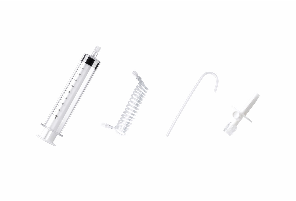
Light changes direction as it passes between liquid and air. A bubble's curved surface redirects that light, making the edge stand out. The syringe barrel curves too, so room lighting, glare, and viewing angle can make the same bubble look different from one side to the other. A faint crescent near the syringe tip may be easier to see from an approved inspection position than through the widest part of the barrel.
A reflection can also mimic a bubble. A change in appearance with viewing angle is a clue, not proof: the appearance of a real bubble can change with the angle too. If the distinction remains unclear, treat the fluid path as unverified rather than assuming the mark is harmless.
A bubble may remain near the outlet or cling to an internal surface rather than float through the center of the barrel. Connectors and valves can make small pockets harder to see. That is why an inspection should follow the fluid route: the syringe, its tip, the connectors, the tubing, and the patient-side components specified in the device instructions. On a dual-syringe injector, a clear contrast syringe does not establish that the saline side is ready.
| Where air is noticed | What may explain it | What to verify |
|---|---|---|
| In the barrel during filling | Air may have entered through the fill path, or remained as liquid entered the syringe. | Verify the filling procedure and inspect the barrel and outlet before proceeding. |
| At the syringe tip after filling | Air can remain in the narrow outlet even when the wider barrel looks clear. | Inspect the outlet directly rather than relying on the barrel or a fluid indicator. |
| At a connector or valve | A small pocket can remain in a fitting or be introduced when the fluid path is opened. | Verify the connection and follow the applicable priming and inspection instructions. |
| In the saline branch of a dual-syringe system | The saline branch can retain air independently of the contrast branch. | Verify both branches and the shared downstream line as directed. |
| In the tubing after a component change | Opening or replacing a component can invalidate the earlier air check. | Follow the device and facility procedure for re-verifying the affected fluid path. |
Where a bubble is first seen is not necessarily where it entered. A pocket noticed at a connector might have moved from upstream, or it might have been trapped during assembly. Note when it appeared and what changed immediately beforehand. A bubble seen after a syringe or tubing change calls for a different investigation from one repeatedly seen at the same point during initial preparation.
1. Hold the injection. If the patient is not connected, do not connect them while air is visible or the check is incomplete. If the patient is already connected, follow the device and facility procedures before manipulating the fluid path.
2. Use the instructions for use, or IFU, for the exact injector and disposable configuration. Priming means filling the specified fluid path with liquid to displace trapped air. The approved sequence, injector-head position, and controls depend on the system. Do not carry over settings or steps from another model.
3. Reinspect the fluid path after the approved preparation steps. Check the syringe tip, connectors, and downstream tubing, not just the barrel. Where applicable, include both contrast and saline branches. Do not use a patient-connected flush to clear a visible air pocket.
4. Escalate if air persists or the cause is unclear. Follow local procedures for replacing or isolating a suspect component. If injection is already underway and air is noticed, stop the injection and follow the facility's incident response procedure. If air may have entered the patient, alert the clinical team immediately and activate the applicable emergency response.
A useful handoff names exactly what has and has not been checked. "The contrast syringe is filled; the saline branch and patient line still need verification" is more actionable than "The injector is ready." If the handoff occurs before preparation is complete, the incoming operator should verify the fluid path rather than infer readiness from the display or a filled syringe.
No. A fluid-presence indicator, an operator confirmation prompt, and an automatic air detector serve different purposes. A fluid-presence indicator can show that liquid is present without proving that the syringe tip is free of trapped air. A confirmation prompt records an operator's response; it does not inspect the line. An automatic detector monitors the location and conditions specified for that injector. Do not assume it checks every upstream branch, fitting, or syringe outlet.
For a department evaluating a new disposable set, one useful training exercise is to trace the path on the actual approved setup, without a patient connected. Have staff identify what they inspect directly, what the system monitors, and what must be rechecked after a change. The exercise tests something a product brochure cannot: whether staff can tell which parts of the fluid path still need verification.
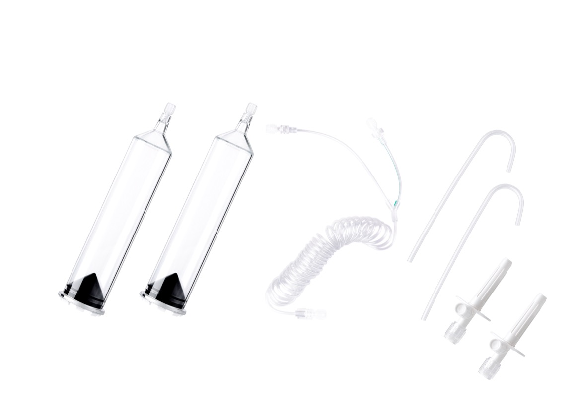
A prospective study of 200 patients undergoing contrast-enhanced chest CT detected incidental venous air on scans in 14 patients, or 7% of that study group. That number describes air detected on chest CT under the study conditions. It is not the percentage of syringes containing bubbles, the rate of symptomatic injury, or a prediction for MRI, angiography, or another CT service. Nor does a scan finding pinpoint the syringe, tubing, or IV connection as the source.
An air embolism occurs when air enters the circulation and can obstruct blood flow. Small incidental CT findings and clinically significant air embolism are not the same outcome. Serious air embolism from intravenous contrast administration is rare, but it can be life-threatening. Neither an incidental finding in a study nor the apparent size of a bubble through the barrel establishes a safe amount of air to inject. The practical rule remains: resolve visible air before proceeding.
If bubbles repeatedly appear in the same place, review the setup systematically. Record the injector model, syringe and tubing part numbers, lot numbers, whether the air appeared on the contrast or saline side, and the point in the preparation sequence when it was first seen. Note whether a connector was opened, a disposable was replaced, or responsibility passed to another operator. If facility policy allows, retain the packaging and implicated components for quality review. A photo taken under facility policy can help document the location without including patient information.
Avoid classifying the event immediately as operator error or a disposable defect. For example, repeated air at one fitting is a reason to review that fitting, the component combination, and the preparation sequence together. The pattern can guide the investigation, but it cannot establish the cause on its own.
A syringe that mounts on an injector or a connector that screws into place is not, by itself, evidence of compatibility. Ask for written documentation that identifies the exact injector model, syringe capacity, disposable part numbers, tubing layout, and labeled operating limits. Include valves, adapters, or other accessories in the review where applicable. For MRI, confirm the labeling relevant to the intended MR environment rather than assuming a set can be transferred from CT. For angiography, verify the intended application and configuration instead of treating a CT workflow as interchangeable.
Inspectability belongs in the purchasing decision too. During an evaluation under normal room lighting, check whether staff can see the syringe outlet and inspect the relevant fittings in the position specified by the instructions. Assess whether labels and lot numbers remain easy to find after the set is unpacked. These checks do not replace technical documentation, but they show whether staff can perform and document the required inspection in practice.
If you source high-pressure syringes, connecting tubing, or related consumables for CT, MRI, or angiography, send MAIWEI INC. your injector model, current disposable part numbers, intended application, and destination market. Request a model-specific compatibility review, applicable instructions for use, labeled operating limits, and product-specific regulatory documentation. If you are investigating recurring bubbles, include the lot number and where in the preparation sequence the air first became visible.
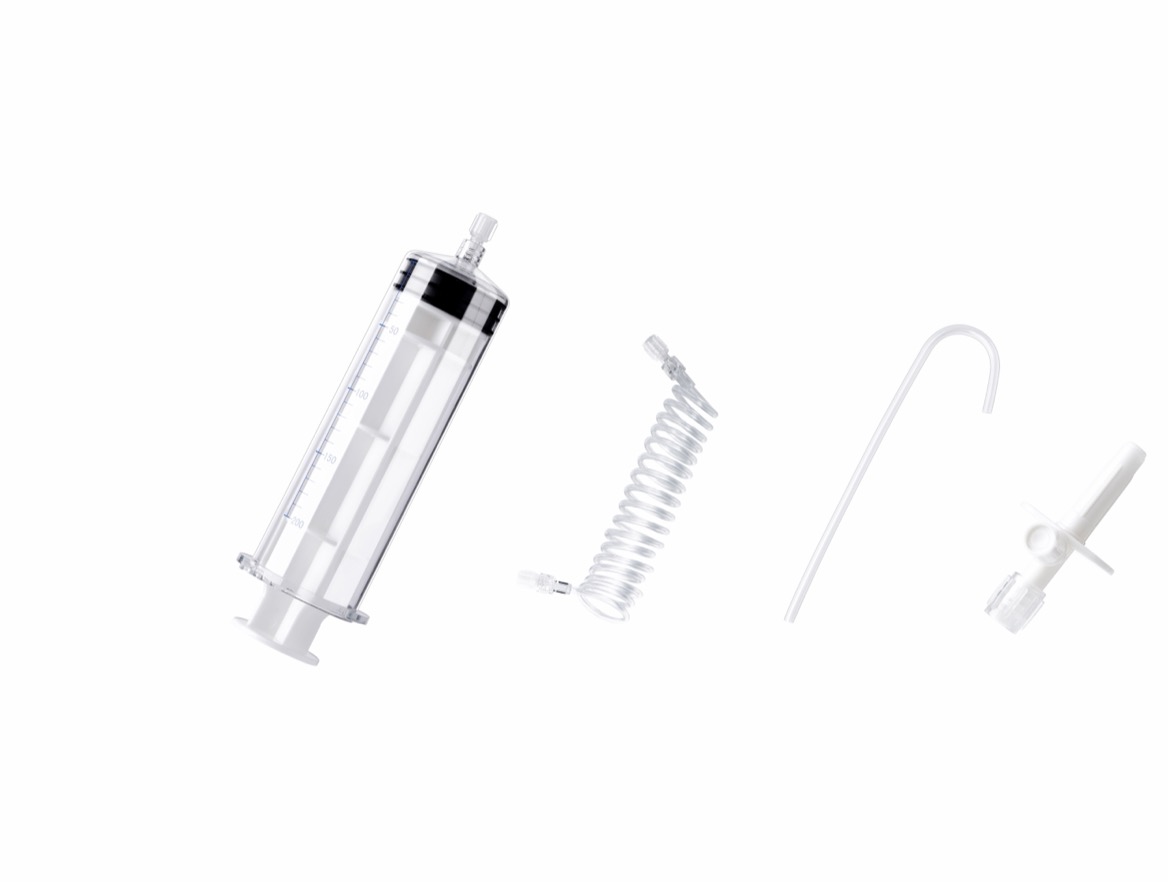
They can appear during preparation, but seeing them during filling does not make them acceptable for injection. Complete the approved air-removal and inspection procedure. If a bubble remains visible or a suspected pocket cannot be ruled out, do not proceed.
Air may remain near the syringe outlet, inside a fitting, or in a branch that was not fully primed. It can also be introduced when a connection is reopened after the original check. Follow the system's instructions for rechecking the affected path before use.
Yes. Fluid presence and absence of trapped air are different observations. A fluid indicator may respond to liquid in the barrel even when a small pocket remains near the outlet. Inspect the areas specified in the operating instructions before confirming readiness.
No. In a dual-syringe setup, the saline branch can feed the same patient line as the contrast branch. Inspect each branch and the common downstream path as required for that configuration. A clear contrast syringe does not verify the saline line.
The objective is the same: clear trapped air before injection. The procedure can differ by injector model, disposable design, and clinical application. Use the instructions for the exact system in service rather than transferring a sequence, setting, or orientation from another injector.
No. Repeated bubbles warrant investigation, but the cause could involve the filling sequence, a connection, an incompatible component combination, or a product issue. Record the model, part numbers, and lot information, then route the case through your facility's quality process and supplier support.
- American College of Radiology. *ACR Manual on Contrast Media*, 2024. See "Safe Injection of Contrast Media" and its discussion of air embolism. [Read the manual]. [geiselmed.dartmouth]
- Pennsylvania Patient Safety Reporting System. *Venous Air Emboli and Automatic Contrast Media Injectors*. Patient-safety advisory covering air entry, staff training, and pre-injection checks. [Read the advisory]. [patientsafety.pa]
- Sodhi KS et al. *Vascular air embolism after contrast administration on 64 row multiple detector computed tomography: A prospective analysis*. *Lung India*, 2015. Prospective study of 200 chest CT patients. [Read the full study]. [pmc.ncbi.nlm.nih]
- Bayer. *MEDRAD Centargo CT Injection System Operation Manual*. Device-specific example of air detection, patient-line inspection, and operator confirmation before arming. [Read the operation manual]. [idsmed.co]
- U.S. Food and Drug Administration. *MAUDE report: MEDRAD Stellant FLEX CT Injection System*. The manufacturer narrative reproduces manual warnings about trapped air, fluid indicators, and operator handoffs. The report is not evidence of event frequency or causality. [Read the FDA record]. [accessdata.fda]